Health
New Imaging Technique Reveals Achilles Tendon Insights in Dancers
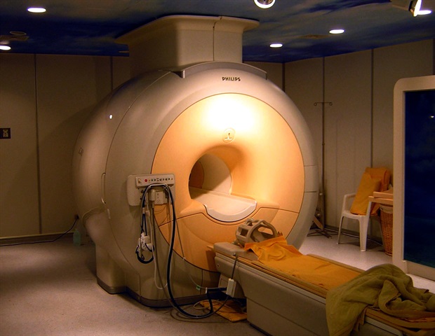
A recent study published in the Journal of Orthopaedic Research introduces a groundbreaking method for assessing the structure and function of the Achilles tendon in professional ballet dancers. The research explores the potential of a noninvasive imaging technique to enhance injury prevention and rehabilitation strategies for athletes and the broader population.
The study utilized a novel approach called multi-echo ultrashort echo time (UTE) magnetic resonance imaging (MRI), which effectively evaluates the collagen and other critical components of the Achilles tendon. This structural assessment was paired with functional evaluations derived from shear wave elastography (SWE) ultrasound, a method that quantifies tendon stiffness.
The findings indicate that professional dancers exhibit greater tendon stiffness compared to their non-dancer counterparts. This observation aligns with previous studies suggesting that repeated physical training can significantly influence tendon characteristics. Specifically, the UTE MRI measurements correlated with the stiffness levels recorded through SWE ultrasound, providing a comprehensive view of tendon health.
Implications for Athletes and Rehabilitation
The authors of the study highlight the significance of integrating UTE and SWE imaging techniques to better understand the structural and functional relationships in tendons. They emphasized that this enhanced assessment of tendon health could lead to improved rehabilitation protocols and more effective injury prevention strategies, particularly for athletes, including professional dancers.
In their conclusion, the researchers state, “These findings highlight the potential of integrating UTE and SWE imaging to investigate tendon structure‐function relationships and adaptations to mechanical loading.” Such insights are crucial, as they may pave the way for tailored rehabilitation programs that consider the unique demands placed on athletes’ bodies.
Future Directions in Research
As the study progresses, further research may refine these imaging techniques and their applications. The potential to apply this method not only to professional athletes but also to the general public opens up new avenues for understanding tendon health and injury risks.
With the study’s promising results, the integration of advanced imaging technologies could transform how sports medicine specialists approach tendon-related injuries, ultimately enhancing the performance and longevity of athletes. As the field evolves, the goal remains clear: to safeguard the health of dancers and athletes through innovative science and informed practices.
For more information, refer to the study by Wiley Horner, A. M., et al. titled “Characterizing Microstructural and Mechanical Properties of Dancer Achilles Tendon Using Ultrashort Echo Time MRI and Shear Wave Elastography Ultrasound,” which will be published in 2025. The study is available at doi.org/10.1002/jor.70075.

Taste Test Reveals Best Mince Pies from Major UK Supermarkets

Baba Vanga Predicts Alien Contact and Global Upheaval in 2026

Astronomers Uncover the Pulsating Mystery of 3I/ATLAS

Ajax Match Abandoned After Fireworks Erupt in Stands

Asia’s Longest Domestic Flights: Some Stretch Over 3,000 Miles

Inmates Sneak in Fried Chicken Takeaways at HMP Wandsworth

Parkdean Resorts Launches Major Black Friday Discounts for 2026 Stays

Former Rockstar Employee Reveals GTA 6 Animations in Leak

Maintain Your HVAC System: Avoid Costly Repairs This Winter

Iconic 90s TV Show House Hits Market for £1.1 Million

Milk Bank Urges Mothers to Donate for Premature Babies’ Health

Alessia Russo Signs Long-Term Deal with Arsenal Ahead of WSL Season

Shoppers Flock to Discounted Neck Pillow on Amazon for Travel Comfort

Museums Body Critiques EHRC Proposals on Gender Facilities

Trump Visits Europe: Business, Politics, or Leisure?
Japanese Teen Sorato Shimizu Breaks U18 100m Record in 10 Seconds

Couple Shares Inspiring Love Story Defying Height Stereotypes

Anglian Water Raises Concerns Over Proposed AI Data Centre
-
 Entertainment2 months ago
Iconic 90s TV Show House Hits Market for £1.1 Million
-
 Lifestyle4 months ago
Milk Bank Urges Mothers to Donate for Premature Babies’ Health
-
 Sports3 months ago
Alessia Russo Signs Long-Term Deal with Arsenal Ahead of WSL Season
-
 Lifestyle4 months ago
Shoppers Flock to Discounted Neck Pillow on Amazon for Travel Comfort
-
 Politics4 months ago
Museums Body Critiques EHRC Proposals on Gender Facilities
-
 Business4 months ago
Trump Visits Europe: Business, Politics, or Leisure?
-
 Lifestyle4 months ago
Japanese Teen Sorato Shimizu Breaks U18 100m Record in 10 Seconds
-
 Politics4 months ago
Couple Shares Inspiring Love Story Defying Height Stereotypes
-
 World4 months ago
Anglian Water Raises Concerns Over Proposed AI Data Centre
-

 Sports4 months ago
Bournemouth Dominates Everton with 3-0 Victory in Premier League Summer Series
-

 World4 months ago
Wreckage of Missing Russian Passenger Plane Discovered in Flames
-

 Lifestyle4 months ago
Shoppers Rave About Roman’s £42 Midi Dress, Calling It ‘Elegant’