Science
Researchers Develop Noninvasive Imaging for Stent Monitoring
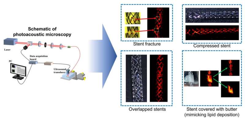
A recent study has demonstrated the potential of using photoacoustic microscopy to monitor stents through the skin, offering a noninvasive alternative to traditional monitoring methods. Each year, approximately 2 million people in the United States receive stents to improve blood flow in narrowed or blocked arteries. This innovative imaging technique could significantly enhance patient safety and comfort during routine evaluations.
Myeongsu Seong, co-lead researcher from Xi’an Jiaotong-Liverpool University, emphasized the importance of monitoring stents for potential issues such as fractures or improper positioning. Conventional monitoring methods often involve invasive procedures or expose patients to radiation. “This inspired us to test the potential of using photoacoustic imaging for monitoring stents through the skin,” Seong stated.
Breakthrough in Photoacoustic Imaging
The findings, published in the journal Optics Letters on July 26, 2025, reveal that photoacoustic microscopy can visualize stents covered with mouse skin under various clinically relevant conditions, including simulated damage and plaque accumulation. Co-lead researcher Sung-Liang Chen from Shanghai Jiao Tong University noted, “While our photoacoustic microscopy results are preliminary, further development could enable frequent, noninvasive monitoring of stent status—without the need for surgical access or X-ray exposure.”
Photoacoustic imaging is a unique technique that detects sound waves generated when materials absorb light and release energy. This method allows for higher-resolution images at greater depths, as sound scatters less than light, making it particularly suitable for monitoring stents.
Advancements in Noninvasive Techniques
In their study, researchers simulated various stent scenarios, including fractures and compression, and even used butter to mimic plaque deposition. Using photoacoustic microscopy at different wavelengths, they successfully imaged these scenarios through excised mouse skin. Seong highlighted a key finding: “We could easily differentiate between the butter we used to mimic a lipid plaque and the stent. Because plaque and stents absorb light differently, using two wavelengths helped us distinguish them.”
The potential applications for this technology are significant. Researchers believe that photoacoustic microscopy could be particularly effective for stents placed in dialysis access sites, typically located just beneath the skin. For stents positioned deeper, such as in the carotid artery, a related approach known as photoacoustic computed tomography may be more effective.
Before this technology can be applied in clinical settings for noninvasive stent monitoring, further in vivo animal tests and preliminary clinical trials will be necessary. Additionally, the imaging system would need to be optimized for various parts of the body.
The development of noninvasive imaging techniques like photoacoustic microscopy represents a promising step forward in the management of stents and cardiac health, potentially improving patient outcomes while reducing risks associated with traditional monitoring methods.
For more information, refer to the study by Siqi Liang et al in Optics Letters (2025). DOI: 10.1364/OL.564778.

LaLiga Thrills: Key Highlights from Recent Fixtures Unveiled

Germany Intensifies Deportations of Afghan and Syrian Migrants

Shoppers Embrace Next’s £36 Knit as Trendy Christmas Alternative

Harbour Energy Announces 100 Job Cuts Amid Industry Challenges

Amy Childs Shares Weight Gain Journey, Emphasizing Self-Love

UK and US Set to Finalize Zero-Tariff Agreement on Pharmaceuticals

Liverpool Activates Backup Plan as Man City Targets Antoine Semenyo

British Father Drowns in Thailand After Being Swept Away by Waves
TorroFX Set to Transform Trading Landscape with Launch on December 1, 2025

Iconic 90s TV Show House Hits Market for £1.1 Million

Milk Bank Urges Mothers to Donate for Premature Babies’ Health

Alessia Russo Signs Long-Term Deal with Arsenal Ahead of WSL Season

Shoppers Flock to Discounted Neck Pillow on Amazon for Travel Comfort

Museums Body Critiques EHRC Proposals on Gender Facilities

Trump Visits Europe: Business, Politics, or Leisure?
Japanese Teen Sorato Shimizu Breaks U18 100m Record in 10 Seconds

Couple Shares Inspiring Love Story Defying Height Stereotypes

Anglian Water Raises Concerns Over Proposed AI Data Centre
-
 Entertainment2 months ago
Iconic 90s TV Show House Hits Market for £1.1 Million
-
 Lifestyle4 months ago
Milk Bank Urges Mothers to Donate for Premature Babies’ Health
-
 Sports3 months ago
Alessia Russo Signs Long-Term Deal with Arsenal Ahead of WSL Season
-
 Lifestyle4 months ago
Shoppers Flock to Discounted Neck Pillow on Amazon for Travel Comfort
-
 Politics4 months ago
Museums Body Critiques EHRC Proposals on Gender Facilities
-
 Business4 months ago
Trump Visits Europe: Business, Politics, or Leisure?
-

 Lifestyle4 months ago
Japanese Teen Sorato Shimizu Breaks U18 100m Record in 10 Seconds
-
 Politics4 months ago
Couple Shares Inspiring Love Story Defying Height Stereotypes
-
 World4 months ago
Anglian Water Raises Concerns Over Proposed AI Data Centre
-

 Sports4 months ago
Bournemouth Dominates Everton with 3-0 Victory in Premier League Summer Series
-

 World4 months ago
Wreckage of Missing Russian Passenger Plane Discovered in Flames
-

 Lifestyle4 months ago
Shoppers Rave About Roman’s £42 Midi Dress, Calling It ‘Elegant’