Science
New Ultrasound Technology Maps Blood Flow in Real Time
Researchers at the Institute Physics for Medicine Paris have developed an innovative ultrasound probe capable of mapping blood flow across entire organs in real time. This breakthrough could significantly enhance the understanding of microcirculation—the flow of blood through the smallest vessels—thus improving the diagnosis of various vascular disorders.
The study, published in Nature Communications, combines 3D ultrasound localization microscopy (ULM) with a multi-lens array method. The team, led by PhD student Nabil Haidour under the guidance of senior author Clement Papadacci, aims to provide a comprehensive view of the circulatory system. Their technique reveals the spatial relationships between macro- and micro-vascular networks, offering insights into the structural and functional organization of organs.
The 3D ULM method localizes intravenously injected microbubbles, achieving a spatial resolution roughly ten times finer than conventional ultrasound. While previously successful in small animal models, visualizing entire organs in larger animals or humans posed challenges due to technological limitations.
To overcome this, the researchers designed a multi-lens array probe consisting of 252 large ultrasound transducer elements. This design increases the probe’s sensitive area while keeping the number of elements relatively low. Each transducer is equipped with an individual acoustic diverging lens, which allows for better signal overlap and coherent image formation.
Whole-Organ Imaging Breakthrough
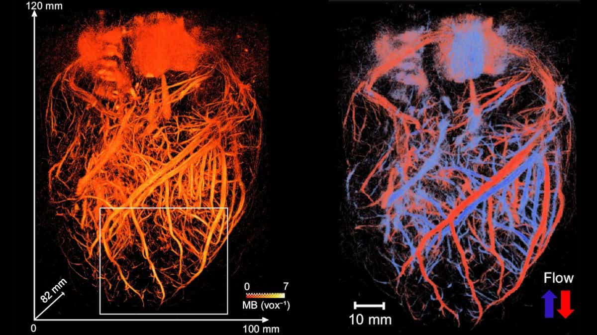
The team validated their method through numerical simulations and experiments before applying it to a whole explanted porcine heart. This organ serves as an effective cardiac model due to its anatomical similarities to human hearts. By perfusing the heart with a microbubble solution, the probe successfully visualized the entire coronary microcirculation network across a large volume of 120 x 100 x 82 mm, achieving a spatial resolution of approximately 125 μm. This method enabled real-time visualization of both large vessels and the finest microcirculation.
The researchers employed a skeletonization algorithm to measure vessel radii, which ranged from about 75 to 600 μm. They also assessed flow dynamics at high temporal resolution, estimating absolute flow velocities from 10 mm/s in smaller vessels to over 300 mm/s in larger ones. The ability to differentiate between arteries and veins based on flow direction marked a significant advancement in vascular imaging.
The team further tested the multi-lens array probe on the kidneys and liver of an anesthetized pig at the Veterinary school of Maison Alfort. The probe was positioned in front of the organs, with electrocardiography synchronizing ultrasound acquisitions to minimize motion artifacts.
The kidney’s vascular network was mapped over a volume of 60 x 80 x 40 mm, achieving a spatial resolution of 147 μm and a maximum absolute flow velocity of around 280 mm/s. In contrast, liver imaging presented more challenges due to various motion factors. Nevertheless, the probe enabled the visualization of a substantial volume of liver vasculature (65 x 100 x 82 mm) with a spatial resolution of 200 μm. The researchers successfully identified the liver’s three blood networks: arterial, venous, and portal veins.
Future Clinical Applications
The combination of whole-organ volumetric imaging with high-resolution vascular quantification addresses significant limitations of existing imaging modalities, such as ultrasound Doppler imaging and CT angiography. While clinical applications of 3D dynamic ULM are yet to be fully demonstrated, Papadacci emphasizes its potential utility in evaluating kidney transplants, coronary microcirculation disorders, stroke, aneurysms, and cancer-related vascular changes.
Looking ahead, the research team anticipates that the technology could soon be applied in human clinical settings. They plan to initiate a clinical trial in early 2026.
This innovative ultrasound technology represents a significant step forward in medical imaging, providing researchers and clinicians with a powerful tool to better understand and treat vascular conditions.

Liverpool Activates Backup Plan as Man City Targets Antoine Semenyo

British Father Drowns in Thailand After Being Swept Away by Waves
TorroFX Set to Transform Trading Landscape with Launch on December 1, 2025

BBC Breakfast’s Sally Nugent Apologizes for News Disruption

Recognize Bowel Cancer Signs During Festive Overindulgence

Marie Helvin Discusses Dating Challenges and Body Confidence at 73

Danny Morrison Reflects on United Ireland, Gerry Adams, and Stormont’s Future

Southampton University and Siemens Collaborate to Advance MEMS Technology

Barcelona’s Deco Highlights Rashford’s Burden at Manchester United

Iconic 90s TV Show House Hits Market for £1.1 Million

Milk Bank Urges Mothers to Donate for Premature Babies’ Health

Alessia Russo Signs Long-Term Deal with Arsenal Ahead of WSL Season

Shoppers Flock to Discounted Neck Pillow on Amazon for Travel Comfort

Museums Body Critiques EHRC Proposals on Gender Facilities

Trump Visits Europe: Business, Politics, or Leisure?
Japanese Teen Sorato Shimizu Breaks U18 100m Record in 10 Seconds

Couple Shares Inspiring Love Story Defying Height Stereotypes

Anglian Water Raises Concerns Over Proposed AI Data Centre
-
 Entertainment2 months ago
Iconic 90s TV Show House Hits Market for £1.1 Million
-
 Lifestyle4 months ago
Milk Bank Urges Mothers to Donate for Premature Babies’ Health
-
 Sports3 months ago
Alessia Russo Signs Long-Term Deal with Arsenal Ahead of WSL Season
-
 Lifestyle4 months ago
Shoppers Flock to Discounted Neck Pillow on Amazon for Travel Comfort
-
 Politics4 months ago
Museums Body Critiques EHRC Proposals on Gender Facilities
-
 Business4 months ago
Trump Visits Europe: Business, Politics, or Leisure?
-

 Lifestyle4 months ago
Japanese Teen Sorato Shimizu Breaks U18 100m Record in 10 Seconds
-
 Politics4 months ago
Couple Shares Inspiring Love Story Defying Height Stereotypes
-
 World4 months ago
Anglian Water Raises Concerns Over Proposed AI Data Centre
-

 Sports4 months ago
Bournemouth Dominates Everton with 3-0 Victory in Premier League Summer Series
-

 World4 months ago
Wreckage of Missing Russian Passenger Plane Discovered in Flames
-

 Lifestyle4 months ago
Shoppers Rave About Roman’s £42 Midi Dress, Calling It ‘Elegant’